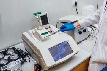
The polymerase chain reaction, or PCR, was originally developed by Kary Mullis, for which he was awarded the Nobel Prize in 1993.
PCR is a laboratory technique that is used to generate large quantities of specified DNA. The polymerase chain reaction produces the selective amplification of a specific type of DNA- fragment for cloning.
The polymerase chain reaction is the cell-free amplification technique, which is used to synthesize multiple identical copies of any DNA of interest.
PCR Principle
The DNA of interest is denatured by the action of restriction endonuclease, which separates the two strands of the DNA. The strand is hybridized with a primer by the process of renaturation. The complex of the resultant duplex is used further for DNA synthesis.
The three steps of denaturation, renaturation, and synthesis take place again and again to produce copies of target DNA.
The PCR Protocol / Technique of polymerase chain Reaction
The following are the essential requirements for PCR procedure like
Reagents
- A target DNA.
- Two primers (synthetic oligonucleotides of 17-30 nucleotides that are complementary to the DNA )
- 4- deoxyribonucleotides (dATP, dTTP, d-GTP, d-CTP )
- A DNA polymerase that can withstand the temperature up to 95 degrees Celsius.
- Reverse transcriptase enzyme
- 30 cycles of PCR are done to acquire the desired DNA sample.
Equipment

- Pcr machines
- Pcr plates
The working mechanism of PCR :
The actual mechanism of PCR involves the following steps, which include the repeated cycles of amplification of target DNA.
Denaturation
This step is also called the melting of the target DNA. In this step, the DNA of interest is subjected to a high-temperature range from 94 to 96 degrees Celsius, resulting in the separation of the two strands of the DNA. Every single strand of the target DNA then acts as the template for DNA synthesis.
Renaturation or annealing
As the temperature of the above mixture is slowly cooled to about 55 degrees Celsius, the primers base pairs with the complementary region flanking the DNA target strands. The two oligonucleotides primers anneal or hybridize to each of the single-stranded DNA. This step operates at a low-temperature range of 40 to 60 degrees Celsius, depending upon the length and sequence of the primers. This process is called annealing or renaturation. The high concentration of primers ensures annealing between each DNA strand and the primers rather than the two strands of DNA.
Synthesis or extension of polymer chains
The synthesis step starts at the 3’-hydroxyl part of each primer. The primers are elongated with the addition of the complementary bands. The synthesis process in PCR is quite comparable to the DNA replication of the leading strand.
The final step is the extension, wherein Taq DNA polymerase (thermophilic bacterium Thermes aquatics) synthesizes the DNA region between, using dNTPs (deoxynucleotides triphosphates) and magnesium ions. The maximum temperature for this step is 72 degrees Celsius.
Like this, all the steps are repeated again and again to give multiple copies of the target DNA.
Other methods include
There are few modifications of the protocol as below.
PCR RFLP Method Protocol
PCR- restriction fragment length polymorphism is also called cleaved amplified polymorphism sequences (CAPS). This technique is famous for its techniques in genetic analysis. PCR-RFLP is applied to detect interspecies in a pool of genomes and analyze their variations.
The method of PCR-RFLP is similar to the normal PCR methods with some addition to it. Mixed type of PCR-RFLP is also executed to detect the genetic variations.
The steps are as follows:
- Digestion of amplicons by internal digestion controls
In this step, the digestion of the relevant amplicon happens via the cleavage process. The strands are recognized by the restriction enzyme. The sequences are prepared for further process. Sometimes, the sequences are not easily recognized; in that case, the internal digestion takes place to extract the amplicon sequences. Plasmids can also be used in this internal digestion process.
- The extracted sequences are then labeled with the fluorescent primers, which can be a fluorescent dye also. This recognizes the RFLP markers for the digestion of sequences. Occasionally, an amplified fragment contains many RFLP markers.
- The resultant mixture is then taken for the electrophoresis and visualization of fragments. These fragments get resolved through electrophoresis. It is done through gel electrophoresis with the help of polyacrylamide or agarose gel. These gels play the role of the molecular sieving matrix.
- The amplification process starts and the fragments get resolved by gel electrophoresis. For analysis in PCR-RFLP, the extracted fragments are usually heated and analyzed by denaturing electrophoresis in a single-stranded state. This is done to determine the fragment sizes to adopting genotyping of microsatellites.
- Thus, amplified DNA sequences are analyzed and a chain of reactions is executed for proper genetic variations.
PCR Amplification Methods
Amplification is an important step in a whole polymerase chain reaction. Without this, PCR is incomplete. The amplification cycle contains three steps:
- Denaturation: the strands are taken into consideration for denaturing it with the help of denaturating enzymes called restriction endonucleases. The denaturation process typically commences at 93-95 degrees Celsius.
- Primer annealing: this step requires a temperature of about 50- 70 degrees Celsius. Here the hybridization of DNA primers happens. The sequences are primed by small DNA sequences. The primers recognize the strands and start the process of annealing.
- DNA synthesis: the third steps mark the beginning of the process of DNA synthesis, which is carried on by hybridizing the sequences of DNA taken into consideration.
The amplification process can be carried out repetitively by varying the temperature of the reaction mixture. This cycle is also called a reaction cycle.
Quantitative PCR Method
This type of PCR is based on the general principle of PCR, which is used to amplify the DNA strands. After amplification, the DNA gets quantified to the target DNA. This is also called real-time quantitative PCR, as it allows scientists to view the increase in the amount of DNA as it is amplified.
This type of PCR is based on the detection and quantitation of a fluorescent reporter, which signals the increase in the direct proportion of the amount of PCR product. The fluorescent probes used here is SYBR which binds to the dsDNA for making it different from others. These fluorescent probes are sequence-specific normally. SYBR green bids to the minor groove of the dsDNA. In this solution, the unbound dye exhibits less fluorescence only. The fluorescence is greatly enhanced when it is bounded with the dsDNA.
Working of Quantitative PCR
- The double-stranded DNA is taken for the process. It is probed by primers called Taq polymerases. A taq man probe is used in the process, which is designed to hybridize an integral part of the PCR region.
- The Taq Man probe has three regions: a short wavelength fluorophore on one end, a sequence-specific to the target DNA, and a quencher at the end. It is seen that as long as the fluorophore and quencher are close to each other, the fluorescence is quenched and fluorescence is observed. But when they are not close, the fluorescence is seen.
- The probe is designed to anneal to the center of the target DNA. When Taq polymerase elongates the second complementary strand during PCR, its 5′ to 3′ endonuclease activity cuts the probe into single nucleotides. This causes the reduction between quencher and fluorophore. Hence, fluorescence is observed. The signal will be detected that is equal to the number of DNA strands synthesized.
PCR Multiplex Method
As the name suggests, multiplex PCR consists of multiple primers, fingerprints, and rapid identification factors. It is used in assisting the determination of a specific cause or species. In this type of PCR, two or more sets of primers are used for different DNA targets in the same PCR reaction. This technique saves considerable time as in one single PCR reaction, multiple specific causes can be analyzed.
The primers used in multiplex PCR are selected carefully based on their annealing temperatures and they seem complementary to each other. The amplicon sizes used in this should be different enough to form different bands on the strand of DNA used. So, multiple bands can be obtained. The bands are then visualized by gel electrophoresis and then the usual PCR reaction happens.
Calculation of PCR
After each cycle, several templates double. So that if one starts with a single dsDNA molecule, after 20 cycles, the number increases to 1x 10⁹. This number is calculated by applying the following formula:
Nf= Ni x 2n
Where Nf = the number of DNA molecules produced by the PCR
Ni= the initial number of molecules (template), and
n = the number of cycles performed
When amplifying a small fragment of the long double-stranded DNA template, the desired blunt-ended target fragments first appear in the third cycle of PCR.
Temperature for PCR
The temperature of PCR varies in its different steps as follows:
- denaturation happens at about 93-95 degree Celsius
- The primer annealing happens at a temperature of about 50 – 70 degrees Celsius. But, the annealing temperature is typically 5 degrees Celsius.
- The DNA synthesis takes place at about 70-75 degrees Celsius.
PCR Method for Bacterial Identification
For bacterial identification, a special rapid PCR is designed by scientists who can identify different species of bacteria. The broad range of primer mixture is first developed using conserved regions of bacterial topoisomerases called genes gurB. This primer design allows the use of DNA amplification further, which is used to produce labeled, single-stranded DNA. These DNA strands are suitable for microarray hybridization. Further, the probes on the microarray are designed to get the designated gene traits. After this, the same cycle of DNA synthesis takes place and PCR reaction concludes.
PCR Cloning Method
Cloning is the backbone of the PCR reaction. PCR cloning is versatile and allows DNA pieces to be placed on the vector’s backbone of choice. The technique has minimal limitations. The copy of DNA forms here by adding restriction sites to the ends of the considered piece of DNA. The plasmid is formed after that. This is known as the cloning method of PCR.
PCR Lamp Method
LAMP stands for loop-mediated isothermal amplification. This method uses 4-6 primers that recognize 6-8 distinct regions of target DNA for the amplification process. At strands, the DNA polymerase initiates the synthesis, and 2 primers form a loop on the strand. The loop structures facilitate the further amplification process followed by renaturation and DNA synthesis.
Qualitative PCR Method
When the PCR method is used to detect the presence or absence of a specific DNA product, it is called qualitative PCR. With this technique, PCR is performed for cloning purposes and the identification of pathogens. It is mainly used for the identification of pathogens.
When is the PCR called successful?
For checking whether the PCR is successful, we can use a gene ladder composed of ethidium bromide, which gives the expression of DNA fragments formed. The size of the DNA fragment produced seems the same as the DNA strands used for amplification.
Why PCR Fails?
Usually, it is seen that the cause of the failure of PCR is the faulty enzyme or reagent used. However, in some times, the irregular primer designs, thermo-cycle parameters, and nonspecific bindings are the cause of the failure of PCR.
The applications of PCR in different areas are as follows.
- PCR in clinical diagnosis.
- In the prenatal diagnosis of inherited disease.
- Diagnosis of retroviral diseases.
- Diagnosis of bacterial diseases.
- In sex determination of embryo.
- In DNA sequencing.
- In a comparative study of genomes.