The ear is one of the important sense organs of the Human Body.
There are a pair of ears on either side of the head.
Though we know our ears help in hearing the sounds. They also help in the maintenance of body balance.
This is why during an ear problem, we suffer from dizziness and vertigo.
The ears perceive the sound from surroundings by converting sound energy (i.e., mechanical energy) into electrical energy, which is carried by auditory neurons to the region of hearing in the brain.
Structure of Ear and its Parts
The ear has many parts that can be classified according to its regions.
Anatomically an ear can be divided into three regions as
- External ear
- Middle ear
- Internal ear.

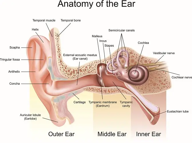
External ear Anatomy
The external ear has three parts like
- Pinna
- auditory canal
- Tympanic membrane
The external ear starts from the part of the ear we see, that protruding part, and is limited in the inner side by the tympanic membrane.
Structures: External, although it may seem devoid of many structures, also has a lot.
A. Pinna
Pinna is the outer helical structure that we can see on each side of the head. It cannot be moved in humans, but in lower animals, it can be moved, like in rabbits, dogs, cows, etc.
The structure of the pinna has cartilage covered up with skin. It is a single piece of yellow cartilage tissue, except for the lower lobule part, which is barely a piece of flesh and skin.
Functions of the pinna: Pinna acts as a condenser; it collects the sound waves from distant sources and channels them onto the tympanic membrane.
It’s an astonishing piece of art of nature that shows perfect engineering. A perfect condensation of sound is achieved through this pinna.
Further, the skin of pinna is used for grafting like nasal ala or of hands. The cartilage is used in reconstruction surgery of the middle ear or nasal bridge.
B. Auditory canal (External Acoustic Canal)
The concha of pinna leads to an ‘S-shaped canal that ends at the tympanic membrane.
In humans, this canal is 24mm long. The outer part is directed upward, backward, and medially and the inner part is directed downward, forward, and medially.
One needs to pull the pinna upward, backward, and laterally to have a look at the tympanic membrane.
The structure of the auditory canal has two parts.
a) The outer one-third of the canal is made up of cartilage. It has skin with hair follicles.
Cerumen and pilosebaceous glands are also seen in this part and are a seat for Staphylococcus bacterial infections too.
b) The inner two-thirds of the canal is made of bone. It is devoid of glands and hair follicles. But cerumen secreted in the outer part can be seen here too.
Functions of the auditory canal
i) It helps to carry the sound waves to the tympanic membrane.
ii) It also helps to avoid the entry of insects and keep the infections and dust away from the inner ear.

C. Tympanic Membrane
The tympanic membrane acts as the barrier between the middle ear and the external ear.
It is 9-10mm in height, 8-9mm in width, and 0.1mm in thickness, placed obliquely.
The tympanic membrane has two basic parts
a) Pars tensa: It is quite stretched with an attachment of the head of the malleus bone.
b) Pars flaccida. It is loose, reddish, and has malleal folds.
Functions of the tympanic membrane
It converts sound energy into mechanical energy in the form of oscillations. These vibrations are transmitted to the inner ear ossicles that, in the course, help in hearing.
Middle Ear
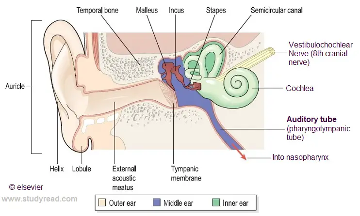
In the above image, the region with a blue shade is in the middle ear.
As we can see, it is the continuation of the tympanic membrane. A closed room, with the tympanic membrane on one side and oval and round windows on another side, the only aperture of this chamber is situated at its base, called the Eustachian tube.
Structures of Middle Ear
The middle ear is a densely packed portion of the ear. It has three small bones called ear ossicles.
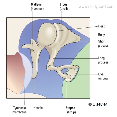
A. Ear ossicles
Three ear ossicles are found in a human, namely-
- Malleus,
- Incus, and
- Stapes.

Malleus
This is a hammer-shaped bone, which is attached to the tympanic membrane (pars tensa part). And the other end is attached to the next ossicle called the incus.
Incus
As seen in the image, it is the next ossicle, which is attached with malleus on one side and with the stapes on the other side.
Stapes
This is the last ossicle and the smallest bone of the human body. It is attached with incus on one side, with the annular ligament on the attic of the middle ear, and on the other side, it is attached to the oval window.
Functions of Ear Ossicles
Ear ossicles play a critical role in hearing. The ear ossicles transfer the vibrations they receive from the tympanic membrane to the round window.
Not only this, but they also act as a safety valve.
They can temporarily shut down hearing if a sound of large amplitude hits the tympanic membrane by losing the annular ligament of stapes.
Moreover, they amplify the sound many times as it reaches the round window.
Two muscles are also attached to these bones, called tensor tympani and stapedius. Stapedius is, no doubt, the smallest muscle of the human body.
B. Auditory tube (Eustachian Tube)
This eustachian tube is a few millimeter-long tubes that connect the middle ear with the pharynx. It is the only vent to the otherwise closed middle ear.
Hence, you will notice that during cold or infection of the respiratory system, your ears may become less audible.
Functions of Eustachian tube
The eustachian tube helps to have a painless hearing.
This auditory tube maintains the middle ear’s air pressure equal to atmospheric pressure as the other side of air is exposed to the atmosphere.
If not for this opening, the pressure will directly act on the tympanic membrane, bending it or tearing it, causing immense pain and deafness.
Other structures in the middle ear, like the mastoid air spaces, the temporal air sinuses, etc., help in hearing in indirect ways.
Internal Ear Anatomy
The internal ear is where the two functions of hearing and balance are processed. It has 2 important parts like
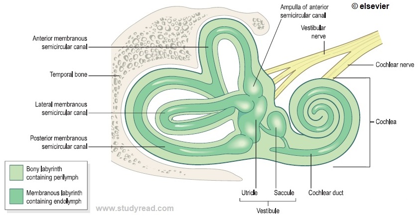
Bony labyrinth
The Bony labyrinth is formed of bone, as the name suggests. It is divided into 3 parts:
A. Vestibule: This is the central chamber of the bony labyrinth. It has two recesses lodging into the utricle. It is the seat of the dynamic body balance.
B. Semilunar Canals: This is the Main seat of body balance in the bony labyrinth. Having calcium carbonate crystals help in the static balance. It has three semicircular canals that act independently. All of them open into vestibule by five openings. All the canals are at a right angle to each other.
C. Cochlea: It is like a snail shell structure with compartments
Functions of the bony labyrinth
It maintains the body balance with the help of the cerebellum, a part of the brain.
Membranous labyrinth.
The membranous labyrinth is mainly formed of membranes, as the name suggests. Parts are:
A. Cochlea: Although the cochlea is a bony spirally coiled tube, the cochlear duct is a Membranous one. The cochlea has three parts, namely
- Scala vestibule
- Scala tympani
- Scala media
Fluids are all around in the internal ear. It covers up all the chambers of the cochlea. The fluid present in scala tympani and vestibule is called perilymph, whereas, that present in the scala media is the endolymph. Both the scala tympani and the vestibule are connected with each other by an opening called Helicotrema. Scala tympani connects with subarachnoid space CSF via the aqueduct of the cochlea.
B. Cochlear duct: It is actually part of the cochlea itself. It consists of three membranes, namely
- Basilar membrane, on which lies the organ of Corti.
- Reissner’s membrane, which separates the organ of Corti from the scala vestibule.
- Striavascularis.
The membranous labyrinth helps in hearing function.
Functions of Internal ear
Among all the functions except visual beauty, others are maintained and completed by the internal ear only.
It has a bony labyrinth, which helps in maintaining body balance via its parts vestibule and semicircular canals.
The Membranous labyrinth has an organ of Corti, which helps in hearing. It has hair cells that move during the vibrations, which are passed by the stapes to the round window, and this opens the sodium-potassium channels among the perilymph and endolymph. The endolymph resembles the intracellular fluid, and the perilymph resembles the extracellular fluid.
The change in concentration of these cations leads to a change in the membrane potential of the Basilar membrane.
This is taken as an impulse by the auditory nerve, the 8th cranial nerve. And we can hear; Simple
The ear is the only organ of sono-reception. It lets us hear the sounds of the world.
Many diseases are caused to the ear, starting from minor diseases like infection, temporary deafness to diseases of significant concern like Chronic suppurative otitis media CSOM, etc.
Fun Facts about the ear
1. Bio-metric tool: The ear is one of the external tools to establish the identity of a person, like a fingerprint or iris of the eye, etc. This fact was first discovered in 1893 by Bertillon. The external features of the ear like ear lobes, presence of piercing, shape, etc. are used for biometric purposes. It is said to be unique in comparison to other biometric traits, due to being less affected by age, unaffected by a change in facial expressions, and also not affected by facial make-up or ornaments that can cover its appearance.
2.Dual function: We assume the ear is meant only for hearing. But, it has two different and essential functions in our body. The inner part of the ear has two functional parts as
a) Choclea: This helps in hearing sound
b) Vestibular system: This helps in the balance of the body.
3. Ear wax: The ears have two glands namely the sebaceous glands and ceruminous glands in the external auditory meatus. Sebaceous glands secrete oils while the ceruminous glands wax. These two secretions are sticky in nature and help to trap dust, insects, and bacteria that try to enter the auditory canal. These secretions along with dead epithelial cells form the ear wax. This wax can sometimes cause pain and deafness until removed mechanically or by the use of warm bicarbonate water.
4. Blood labyrinth barrier: This is the barrier similar to the blood-brain barrier, which separates the inner ear from systemic circulation. Due to this, drugs taken by the systemic route do not reach the inner ear leading to ineffective treatment in case of infections or diseases to the ear. Hence, ear treatment can include other routes of drug administrations.
5. Body balance:
The inner ear vestibular system is the key to the maintenance of the balance of our body. It has 3 semi-circular canals, each helping in sensing one type of movement. The first one helps in senses up-and-down movement. The second canal helps in sensing side-ward movements. The third canal helps in sensing the tilting movements. These canals have fluid and hair cells inside. During movement, the fluid and hair cells move, and the hair cells send these signals to the brain through the acoustic nerve. Our brain uses this information to interpret where we are in space. We tend to get vomiting when this balance system is agitated or disturbed.
6. Hearing loss: With age, there is a loss of sensitivity to a higher frequency. Deafness is caused due to infections, immune disorders, ototoxicity by drugs, and antibiotics. Bacterial and viral infections are prime causes, while aminoglycoside antibiotics damage hair cells.
7. Hearing: The energy of sound waves is converted into neuronal energy, and a signal is conducted to the brain. When the sound waves reach the tympanic membrane, a series of movements is set down, which leads to waves in fluids of the cochlea. This stimulates sensory cells that send auditory stimuli to the brain.
8. Hearing frequency: Sound waves travel in the form of waves and show wave nature i.e., have both frequency and also wavelength. Human ears can hear a sound frequency in the range of 20 to 20 K hertz.
9. Ear in the fetus: A baby develops ears by the 16th week and is able to hear. The ears erect out and stand on the sides of the head at this period.
