The cow’s eye is an organ of incredible complexity.
It is a small and delicate organ but allows a cow to see its surroundings without conscious effort.
The eye’s automatic focusing system is faster than any camera, enabling one to see both in starlight and the brightest sunlight.
Each part of the eye works together to provide information to the brain, which processes it instantaneously to give a perfect image.
Cow eyes are similar to human eyes in some aspects. Both humans and cows have a cornea in the front and a lens behind it.
Studying the cow’s anatomy can teach how the human eye works and functions.
Cow’s Eye External Anatomy Parts


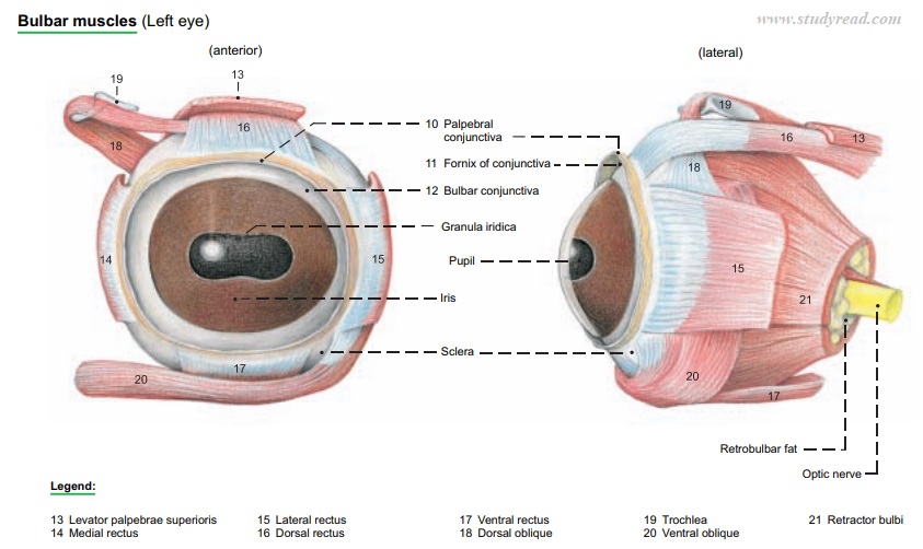
Sclera and cornea.
It is a thick, white protective layer forming a cover on the outer side of the eye body.
This sclera appears as the large mass of grey tissue surrounding a cow’s eye.
The fibrous tunic comprises the sclera and the transparent cornea.
The sclera encloses the greater part of the bulb in its dense white connective tissue.
These parts join at the corneal limbus, which is the border between the sclera and cornea.
The cornea
This is a clear, transparent, epithelial extension of the sclera.
It forms the anterior portion of the sclera section and allows light rays to pass on to the lens.
This cornea is convex towards the anterior and helps to refract (bend) the light rays so as to focus on the retina.
It helps to protect the inner layers of the eye from damage
Conjunctiva
It is a thin mucous membrane that covers the anterior surface of the eyeball.
One can clearly distinguish it from the anterior sclera.
Optic Nerve
The nerve, with a diameter of around 3mm, is placed posteriorly and medially in the eye socket, which works as a mediator for signals.
This optic nerve is a bundle of nerve fibers that connects the eye to the brain.
It carries the electrical signals from the retina to the brain. It helps the brain to interpret visual images and create mental images.
Externally attached eye muscles.
The eyeball’s posterior surface has six muscles connecting it to the cow skull’s bony orbit.
The lens’ shape can be changed by these external muscles linked to the eye, allowing it to concentrate on objects at different distances.
The lens changes shape as these muscles are tightened (contraction) or relaxed (relaxation), allowing the eyes to concentrate on close or far things.
This is called accommodation.
Accessory Organs
The eyelids, conjunctiva, lacrimal apparatus, the cone of striated bulbar muscles with their fasciae, and the nerves are all the accessory organs of the eye.
The Upper and Lower Eyelids
The upper and lower eyelids (palpebra superior and inferior) are made of an outer layer of hair skin, a middle fibromuscular layer, and the palpebral conjunctiva.
The fibrous part of the middle layer is attached to the osseous orbital margin and increases in density towards the free border to form the tarsus, which contains the tarsal glands.
Conjunctiva
The palpebral conjunctiva joins the bulbar conjunctiva at the fornix and ends at the limbus of the cornea. The third eyelid is made up of a conjunctival fold in the medial angle that encloses the T-shaped outer end of the third eyelid cartilage.
The deep part of the cartilage is surrounded by the gland of the third eyelid, larger than in the horse, extending about 5 cm straight back into the fat medial to the eyeball and discharging tears through orifices on the bulbar side of the third lid.
Lacrimal Apparatus
The lacrimal apparatus lies in the dorsolateral quadrant of the orbit, with the broad dorsal part under the root of the zygomatic proc. And a long thin tail that extends around the lateral margin of the orbit.
The lacrimal ducts connect the apparatus’s ventral end with orifices in the lateral fornix.
The accessory lacrimal gland of the third eyelid is the largest. Tears collect in the lacrimal lake in the medial angle anterior to the third eyelid around the lacrimal caruncle.
The upper and lower lacrimal puncta and the lacrimal canaliculi drain into the lacrimal sac.
The nasolacrimal duct drains this to the nasolacrimal orifice, which is hidden on the medioventral surface of the alar fold.
Cow’s Eye Internal Anatomy Parts
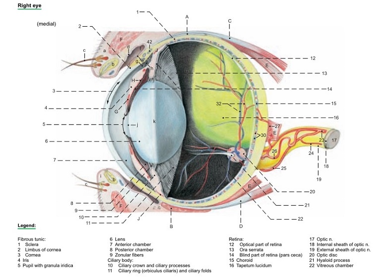
The internal structures of the cow’s eye comprise the following:
Anterior cavity (consists of anterior and posterior chambers)
- The aqueous humor (a watery liquid) is contained in the space between the lens/suspensory ligaments and the cornea.
- The anterior part is the space between the iris and the cornea. The posterior chamber is the space between the lens and the iris.
Posterior Cavity
- The posterior cavity is the cavity formed between the lens and the retina.
- This posterior cavity is filled with vitreous humor liquid that extends between the lens and the retina.
- The vitreous humor present is a transparent gel-like substance that enables the seamless passage of light from the aperture to the retina.
- The stroma is a network that holds a cell-free jelly, the water content of which determines the intraocular pressure.
Pupil
- There is an aperture in the middle of the iris called a pupil. The pupil of a cow is oval.
- A strong, transparent cover (cornea) present protects the iris and pupil of the cow’s eye.
- As the light passes through the cornea, it bends. Pupils serve as the initial stage in creating a retinal image of an object.
Iris
- The sheet of round-shaped smooth muscle found in the interior of the eye is called the iris.
- It is easily distinguishable as it is the colored part of the eye.
- A cow’s iris is brown colored, and a clear fluid called aqueous humor is present around the cornea that helps to maintain its circular shape.
Ciliary Body and Muscle
- A series of radiating folds that appear posterior to the iris of the cow’s eye is called the ciliary body.
- These suspensory ligaments are attached to the anterior part of the eye with a thicker section of the choroid.
This is called the ciliary body.
Suspensory Ligaments
- The ligaments connect the eye lens to the ciliary body and suspend it in position.
- These ligaments help in keeping the eyeball in its usual position such that the balls stay in an upward, forward position in orbit.
- They also prevent the downward movement of the eyeball.
Lens
The lens is Spherical and has a clear transparent structure located behind the cornea.
- The lens is responsible for focusing the light rays entering the cow’s eye onto the retina.
- The eyeball has some blood vessels that supply blood to the cow’s eye for normal functioning.
- This structure, when preserved, turns opaque.
Retina
- This is the innermost layer and is present below the choroid membrane.
- It is found only in the posterior cavity of the eye socket.
- This retina is thin, delicate, and a cream-colored sheet of tissue that forms the screen for image formation.
- In the cone cells of the cow’s retinas, there are only two types of color receptors. Cows, like the majority of non-primate land mammals, are dichromatic.
- The fovea centralis has two to three rods per cone, but the optic papilla has five to six.
- Long-wavelength colors (yellow, orange, and red) are easier for cattle to identify than shorter-wavelength hues (blue, grey, and green).
- Calves can distinguish between long (red) and short (blue) or medium (green) wavelengths, but only to a limited extent between short and medium wavelengths.
- Under red lights, they also approach handlers more quickly. A cow has good color awareness; it does not compare to humans or sheep.
Optic Disc
- A single point of attachment of the retina to the optic nerve is called an optic disc.
- This attachment is also called a blind spot.
When cow edges of their bodies form a blind hole behind them as they elevate their heads, they can see in all directions without rotating their heads because they have a panoramic vision.
Still, binocular vision is limited to around 25 to 50 degrees in front of them.
Choroid
- The eye’s middle layer and posterior portion are always pigmented and highly vascularized.
- It is seen internally by peeling away a portion of the retina from the eye portion.
Tapetum Lucidium
This is one of the exclusive structures found only in cows and other mammals.
It appears as an iridescent silver-blue pigmented portion of the choroid.
Its primary function is to reflect light in the posterior cavity for increased light absorption efficiency under low-light conditions.
Differences Between a Cow’s Eye and a Human Eye
| Characteristics | Cow’s Eye | Human’s Eye |
|---|---|---|
| Size | The average cow eyeball has a radius of a little over 1/2 inch (15mm) and a diameter of 1.2 inches (30mm) | The diameter of the human eyeball varies, but on average, it is roughly 1 inch (24mm). |
| Pupil shape | The shape of the pupil is oval in cows | The form of the pupil is round in humans. |
| Iris Color | The color of Iris is brown. | Iris color can be of a variety of colors. |
| Presence of tapetum lucidium | It is present in the cow’s eye. | Iris color can be in a variety of colors. |
| Rods and cones | fewer in cows. | The number of rods and cones is numerous. |
| External eye muscles | Cows have four muscles and can only see in four directions: up, down, left, and right. | Humans have six eye muscles that enable them to move their eyes in six directions: up, down, left, right, clockwise, and counter-clockwise. |
Conclusion
The cow’s eye has a very close resemblance to the human’s eye.
Due to this fact, the dissection of a cow’s eye is easy to execute and understand.
Frequently asked questions and answers.
How does the cow eye work?
When the light falls on a cow’s eye, it is passed through the pupil to the retina. The light rays are perceived by the optic nerve, which carries the information to the brain for image processing.
How is the cow eye similar to the human eye?
The cow eye is similar to the human eye in terms of the anatomy and physiology of the eyes.